

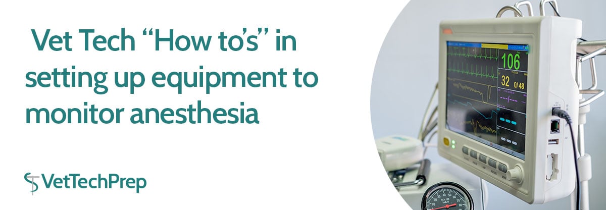
As a vet tech, you will be responsible for setting up the equipment so that you can monitor your patients. Below, we will cover some of the key pieces of anesthetic monitoring equipment and how to set them up. However, there may be differences in equipment types and availability from hospital to hospital, so make sure to ask your supervisor for thorough training on how to set up equipment at your hospital.

As important as it is to know how to set up your anesthetic monitoring equipment, don’t forget the most valuable part of the monitoring process - you! There are many vital parameters that need to be assessed manually such as anesthetic depth, mucus membrane color, capillary refill time, pulse quality, pain assessment, cardiothoracic auscultation, respiratory rhythm and proper function of equipment.
Additionally, many vitals measured by equipment should also be measured manually, such as heart rate, respiratory rate and temperature, among others. Make sure to always manually check and assess your patient - never just rely on the equipment!
Commonly used Monitoring Equipment in Small Animal Practice
-
ECG
-
Typically consists of 3 leads that are color-coded. Leads are clipped to patient’s skin and alcohol is applied over the area; the signal is transmitted to a monitor and is displayed as heart rate and rhythm.
-
Traditionally, the red clip gets attached near the inguinal fold of the left hind leg. The white clip is attached near the axillary area of the right front leg. The black clip is attached near the axillary area of the left front leg.
-
An example of the placement of ECG leads is shown below:

-
Pulse Oximetry
-
Usually consists of a probe that gets placed on an area that does not have hair or pigment (if possible). The signal is transmitted to the monitor and is displayed as SpO2, which is defined as the percent of hemoglobin saturated with oxygen. Pulse rate is also measured and displayed on the screen.
-
An example of placement of the SpO2 probe is shown below:

-
Indirect blood pressure monitoring
-
There are two commonly used types of indirect blood pressure monitoring equipment. Both methods require appropriate sizing and measurement of a cuff. If using a forelimb, usually the cuff is applied at the level of the radius; if using a hindlimb, the cuff is usually applied at the level of the tibia. Key differences between the methods are listed below.
-
1) Oscillometric Method
-
Measures and displays systolic, diastolic and MAP (mean arterial pressure) on the monitor screen
-
You do not have to manually inflate or deflate the cuff - it is done automatically by the machine
-
2) Doppler Method
-
An area distal to the cuff (usually proximal to the metacarpal or metatarsal pads) is shaved
-
Ultrasound coupling gel is applied to the shaved area, then the Doppler crystal is placed on this area
-
A bulb sphygmomanometer is used to manually inflate the cuff
-
Measures systolic blood pressure only
-
An example of the Doppler crystal placement is shown below:

-
Capnography
-
Usually consists of an attachment or adaptor that gets placed on the end of the ET tube. It measures and displays ETCO2 and respiratory rate.
-
Temperature
-
Usually measured rectally via a thermometer or rectal probe. This measurement of core body temperature is displayed on the thermometer or on the monitor. Alternatively, some practices may use an esophageal probe instead.
-
An example of manual measurement of temperature using a rectal thermometer is shown below (note: it would be ideal to use a thermometer probe cover):

The monitoring equipment covered in this article represents the most commonly used pieces of equipment in small animal general practice. Other more invasive types of monitoring equipment include (but are not limited to) direct blood pressure monitoring and arterial blood gas measurements.

Want to learn more? For more information, check out some of the articles listed in the Reference section below!
References:
-
Anesthetic Monitoring. VetTechPrep.com
-
Anesthesia for the Pet Practitioner. Banfield Pet Hospital.
-
Anesthetic Monitoring - What Should You Look For? Kortright Animal Hospital.
-
Burns, Susan. “Anesthetic monitoring savvy.” DVM 360.
-
Monitoring the Anesthetized Patient. Rural Area Vet.
-
Pulse Oximetry. VetGirl.