

February is Dental Month, and we have an inforgraphic of the anatomy of the tooth.
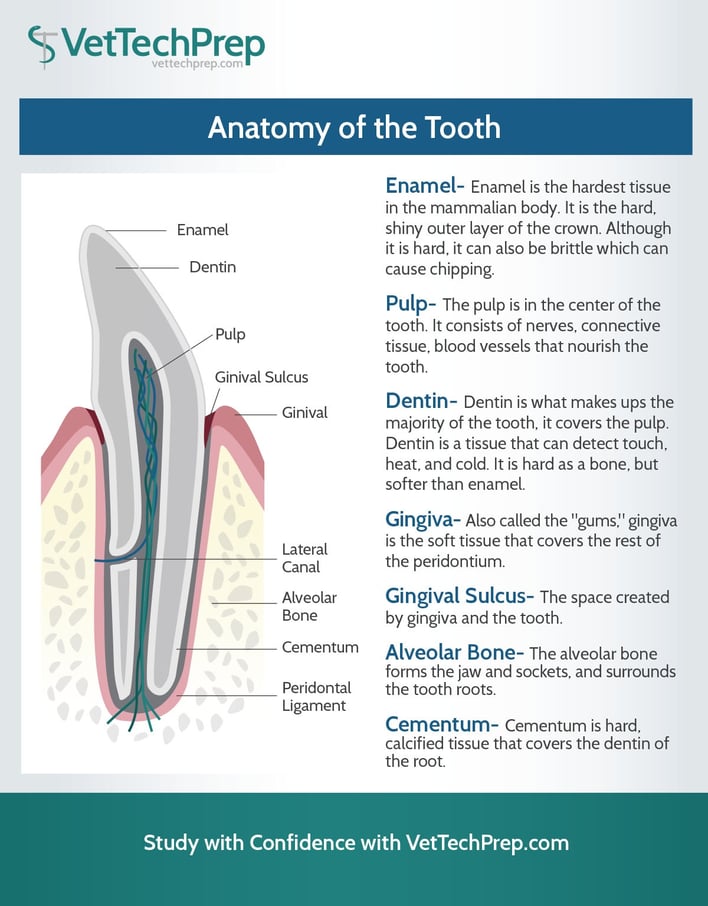
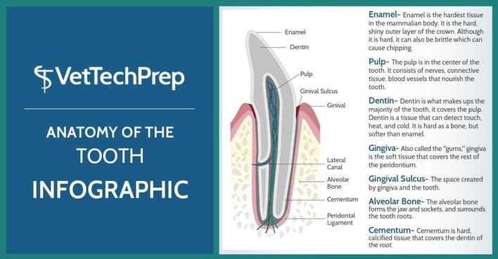
- Enamel is the hardest tissue in the mammalian body. It is the hard, shiny outer layer of the crown. Although it is hard, it can also be brittle which can cause chipping.
- The pulp is in the center of the tooth. It consists of nerves, connective tissue, blood vessels that nourish the tooth.
- Dentin is what makes ups the majority of the tooth, it covers the pulp. Dentin is a tissue that can detect touch, heat, and cold. It is hard as a bone, but softer than enamel.
- Gingiva, also called the "gums," gingiva is the soft tissue that covers the rest of the peridontium.
- Gingival Sulcus, the space created by gingiva and the tooth.
- Alveolar Bone, The alveolar bone forms the jaw and sockets, and surrounds the tooth roots.
- Cementum is hard, calcified tissue that covers the dentin of the root.
- The Peridontal Ligament helps hold the tooth in the socket. It attaches to the cementum of the tooth and the alveolar bone.
- Lateral Canal is a very small channel that connects the root pulp to the periodontal tissue where small blood vessels run.
Be sure to check out our other fact posts.

in
Fact of the Day,
Teeth
0 Comments